Anatomical Name Of Lower Back Muscles - Back Muscles in a Nutshell - Anatomy Tutorial - YouTube
Anatomical Name Of Lower Back Muscles - Back Muscles in a Nutshell - Anatomy Tutorial - YouTube. The pelvic floor muscles also help increase this pressure, which provides stability to the spine and trunk. Related posts of muscle names of lower back muscle anatomy multiple choice. The quadratus lumborum muscles (orange, in the image above) are found in the lower back (also called the lumbar area). Muscle anatomy ankle 12 photos of the muscle anatomy ankle ankle anatomy muscle. Extrinsic and intrinsic.the back functions are many, such as to house and protect the spinal cord, hold the body and head upright, and adjust the movements of the upper and lower limbs.
ads/bitcoin1.txt
All the extrinsic back muscles are innervated by the ventral (anterior) rami of the cervical spinal nerves , except for the trapezius muscle which receives its supply from the accessory nerve (cn xi) . The spine is composed of 33 bones called vertebrae, which stack together to form the spinal canal. Extrinsic and intrinsic.the back functions are many, such as to house and protect the spinal cord, hold the body and head upright, and adjust the movements of the upper and lower limbs. Anatomynote.com found muscles of lower back diagram from plenty of anatomical pictures on the internet. By the way, have you heard about the myth of.
Back Workouts for Women: 4 Ways to Build Your Back by ... from www.bodybuilding.com The latissimus dorsi is the largest and most powerful of your back muscles. The quadratus lumborum muscles (orange, in the image above) are found in the lower back (also called the lumbar area). The lats are attached to the upper end of the humerus with fibers running down in a fan down the vertebral column and pelvic girdle. The lumbar spine is the lower back that begins below the last thoracic vertebra (t12) and ends at the top of the sacral spine, or sacrum (s1). They help to bend the back to one side or the other. Rotate head to the same side. In order to best help your clientele, it's important for coaches to understand the muscles of the back, what can cause back pain. The muscles of the back with the surface (trapezius, latissimus dorsi, thoracolumbar fascia, deltoid) and intermediate layers (serrated muscles, external and internal oblique muscle).
Related posts of muscles of the lower back and hip diagram muscle anatomy ankle.
ads/bitcoin2.txt
We are pleased to provide you with the picture named muscles of lower back diagram.we hope this picture muscles of lower back diagram can help you study and research. Anatomical name of lower back muscles : This curve, called lordosis, helps to: The lumbar spine is the lower back that begins below the last thoracic vertebra (t12) and ends at the top of the sacral spine, or sacrum (s1). The quick answer to this question is the muscles of the lower back are the multifidus, longissimus, spinalis, and quadratus lumborum. In order to best help your clientele, it's important for coaches to understand the muscles of the back, what can cause back pain. The vertebral column of the lower back includes the five lumbar vertebrae, the sacrum, and the coccyx. The back is the body region between the neck and the gluteal regions. Muscle anatomy ankle 12 photos of the muscle anatomy ankle ankle anatomy muscle. It comprises the vertebral column (spine) and two compartments of back muscles; Leaning back to straight vertical and all points in between. It consists of nerves that carry messages to and. The lower part of the trapezius ascends and depresses the scapula, while the transverse or middle region of the trapezius is what retracts the.
All the extrinsic back muscles are innervated by the ventral (anterior) rami of the cervical spinal nerves , except for the trapezius muscle which receives its supply from the accessory nerve (cn xi) . The lumbar spine is the lower back that begins below the last thoracic vertebra (t12) and ends at the top of the sacral spine, or sacrum (s1). Rotate head to the same side. To build the back optimally, you should know the major muscles, their actions, and which exercises build muscles best. The muscles of the back with the surface (trapezius, latissimus dorsi, thoracolumbar fascia, deltoid) and intermediate layers (serrated muscles, external and internal oblique muscle).
Rezultat imagine pentru leg muscle model labeled ... from i.pinimg.com The back muscles represented on an anatomical chart and on a schematic view of the origin and insertion of the proper muscles of the back (iliocostal muscle of. Bones of the pelvis and lower back the bones of the pelvis and lower back work together to support the body's weight, anchor the abdominal and hip muscles, and protect the delicate vital organs of the vertebral and abdominopelvic cavities. Serratus posterior superior and serratus posterior inferior muscles. The back anatomy includes the latissimus dorsi, trapezius, erector spinae, rhomboid, and the teres major. The vertebral column of the lower back includes the five lumbar vertebrae, the sacrum, and the coccyx. Located at the front of your body, the flexors. By the way, have you heard about the myth of. This curve, called lordosis, helps to:
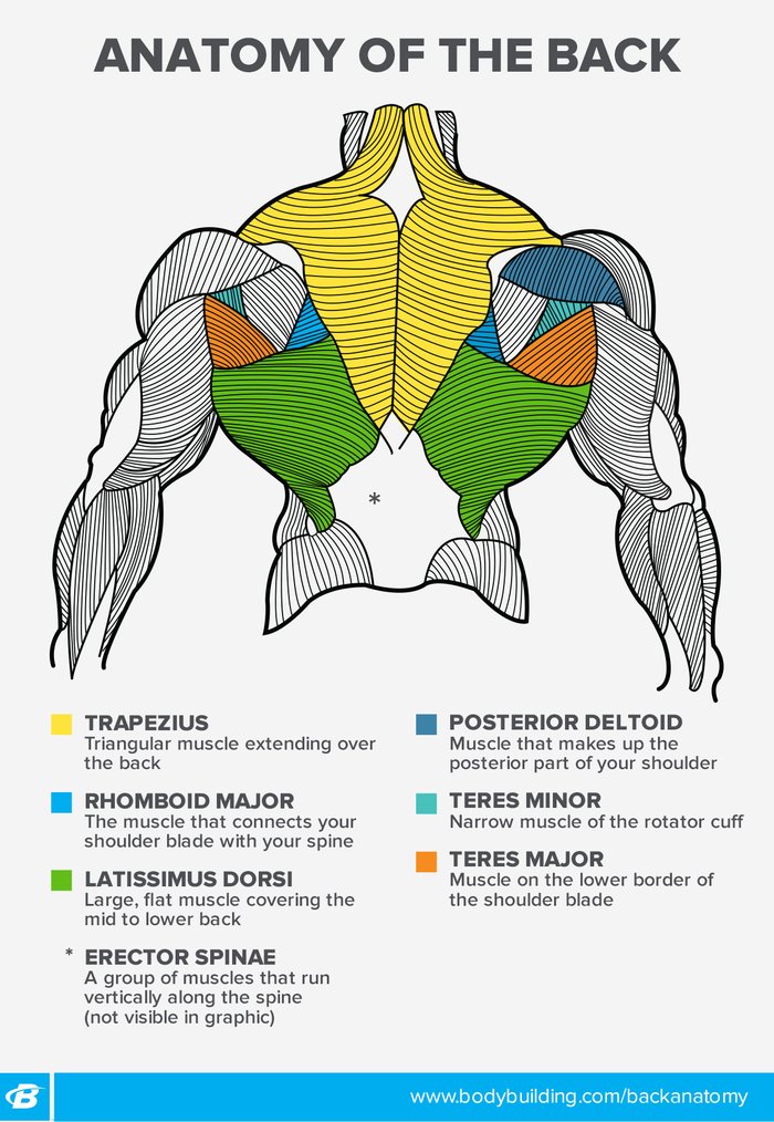
The back anatomy includes the latissimus dorsi, trapezius, erector spinae, rhomboid, and the teres major.
ads/bitcoin2.txt
The back muscles represented on an anatomical chart and on a schematic view of the origin and insertion of the proper muscles of the back (iliocostal muscle of. For more anatomy content please follow us and visit our website: The two main muscle groups involved in back function are: Scientific studies using sophisticated tools such as electromyography (emg) and magnetic resonance imaging (mri) show us how these muscles work. Serratus posterior superior and serratus posterior inferior muscles. Muscle anatomy multiple choice 12 photos of the muscle anatomy multiple choice anatomy muscular system multiple choice questions, muscle anatomy multiple choice questions, muscle anatomy multiple choice quiz, human muscles, anatomy muscular system multiple choice questions, muscle anatomy multiple choice questions. The extensors, which include the many muscles that attach to the spine and work together to hold your back straight while enabling you to extend it. It comprises the vertebral column (spine) and two compartments of back muscles; It is composed of trapezius, latissimus dorsi, rhomboid major, rhomboid minor and levator scapulae. Each lumbar spinal level is numbered from top to bottom—l1 through l5, or l6. The lordotic curve your lower back (lumbar spine) is the anatomic region between your lowest rib and the upper part of the buttock. These muscles provide posture and stability to the body by holding the vertebral column erect and adjusting the position of the body to maintain balance. It starts all the way down in your lower back, climbs up to the middle of your back, and stretches out into your shoulder.
The back anatomy includes the latissimus dorsi, trapezius, erector spinae, rhomboid, and the teres major. Muscle anatomy multiple choice 12 photos of the muscle anatomy multiple choice anatomy muscular system multiple choice questions, muscle anatomy multiple choice questions, muscle anatomy multiple choice quiz, human muscles, anatomy muscular system multiple choice questions, muscle anatomy multiple choice questions. Each lumbar spinal level is numbered from top to bottom—l1 through l5, or l6. The lats are attached to the upper end of the humerus with fibers running down in a fan down the vertebral column and pelvic girdle. The extensors, which include the many muscles that attach to the spine and work together to hold your back straight while enabling you to extend it.
Muscle Chart: Anatomical Muscle Chart - SteroidsLive from www.steroidslive.com We are pleased to provide you with the picture named muscles of lower back diagram.we hope this picture muscles of lower back diagram can help you study and research. Anatomical name of lower back muscles : Intermediate extrinsic muscles of the back: In the meanwhile, your hip flexors, quadriceps and lumbar muscles remain tight to keep you in an upright position. Anatomynote.com found muscles of lower back diagram from plenty of anatomical pictures on the internet. The latissimus dorsi is the largest and most powerful of your back muscles. Related posts of muscle names of lower back muscle anatomy multiple choice. It starts all the way down in your lower back, climbs up to the middle of your back, and stretches out into your shoulder.
Low back pain treatments manchester osteopathy / the lower part of the trapezius ascends and depresses the scapula, while the transverse or middle region of the trapezius is what retracts the.
ads/bitcoin2.txt
The muscles of the lower back, including the erector spinae and quadratus lumborum muscles, contract to extend and laterally bend the vertebral column. They help to bend the back to one side or the other. Rotate head to the same side. It consists of nerves that carry messages to and. The pelvic floor muscles also help increase this pressure, which provides stability to the spine and trunk. Anatomical name of lower back muscles : The lower part of the trapezius ascends and depresses the scapula, while the transverse or middle region of the trapezius is what retracts the. The back anatomy includes the latissimus dorsi, trapezius, erector spinae, rhomboid, and the teres major. All the extrinsic back muscles are innervated by the ventral (anterior) rami of the cervical spinal nerves , except for the trapezius muscle which receives its supply from the accessory nerve (cn xi) . Bones of the pelvis and lower back the bones of the pelvis and lower back work together to support the body's weight, anchor the abdominal and hip muscles, and protect the delicate vital organs of the vertebral and abdominopelvic cavities. The back muscles represented on an anatomical chart and on a schematic view of the origin and insertion of the proper muscles of the back (iliocostal muscle of. Leaning back to straight vertical and all points in between. Located at the front of your body, the flexors.
ads/bitcoin3.txt
ads/bitcoin4.txt
ads/bitcoin5.txt
0 Response to "Anatomical Name Of Lower Back Muscles - Back Muscles in a Nutshell - Anatomy Tutorial - YouTube"


0 Response to "Anatomical Name Of Lower Back Muscles - Back Muscles in a Nutshell - Anatomy Tutorial - YouTube"
Post a Comment